Loculated Pleural Effusion Radiology Ct / Malignant Pleural Effusion - Pulmonology Advisor : (a) axial, (b) coronal, and (c) sagittal images show a large amount of uniform material of fluid attenuation filling much of the right.
Loculated Pleural Effusion Radiology Ct / Malignant Pleural Effusion - Pulmonology Advisor : (a) axial, (b) coronal, and (c) sagittal images show a large amount of uniform material of fluid attenuation filling much of the right.. 19 28 films show rounded density representing a rounded atelectasis in the right. Tuberculosis (mtb) is required in cases of tuberculous pleural effusion (tbpe) for confirming diagnosis and successful therapy. In loculated parapneumonic effusions computed tomography (ct). Ct scans for pleural effusion should be performed with contrast enhancement of the pleura and before complete drainage of pleural fluid. Margins, scalloped borders, and relatively homogeneous attenuation.
Pleural effusion is an accumulation of fluid in the pleural cavity between the lining of the lungs and the thoracic cavity (i.e., the visceral and parietal pleurae). In loculated parapneumonic effusions computed tomography (ct). Primary pleural angiosarcoma as a mimicker of mesothelioma. Large pleural effusions, s/p thoracentesis with pleural fluid suggestive of transudative process. The lungs and the chest cavity both have a lining that consists of pleura, which is a thin membrane.
Pleural diseases chest radiology part1 from image.slidesharecdn.com Learn about different types of pleural effusions, including symptoms, causes, and the pleura is a thin membrane that lines the surface of your lungs and the inside of your chest wall. Improved after thoracentesis and diuresis. In healthy lungs, these membranes ensure that a small amount of liquid is present between the lungs. Light's criteria can be used to determine the type of a patient's pleural effusion and thus its etiology. (ct scan findings consistent with pleural metastasis and. Meaning of loculated pleural effusion medical term. Pleural effusion, the pathological accumulation of fluid in the pleural space, is very common. Primary pleural angiosarcoma as a mimicker of mesothelioma.
Most likely secondary to left ventricular diastolic dysfunction.
Margins, scalloped borders, and relatively homogeneous attenuation. Pleural effusion is an accumulation of fluid in the pleural cavity between the lining of the lungs and the thoracic cavity (i.e., the visceral and parietal pleurae). Obliteration of left costophrenic angle with a wide pleural based dome shaped opacity projecting into the lung noted tracking along the cardiophrenic angle and lateral chest wall suggestive of loculated pleural effusion, however the. Improved after thoracentesis and diuresis. Pleural effusions are characterized on ct by attenuation values between those of water (0 hounsfield units hu. There is smooth thickening of the parietal pleura (arrowhead). Ct of the thorax ± abdomen: Meaning of loculated pleural effusion medical term. Loculated effusions on ct scans tend to have a lenticular shape with smooth. Pleural effusion | radiology key. E7.4 ct of pleural effusion. Loculated pleural effusion radiology case. (a) axial ct scan reveals a left pleural effusion in a patient presenting with back pain.
2 1 right pleural effusion blunting the right costophrenic angle extending posteriorly with some fluid tracking into the major fissure seen on the lateral exam. Meaning of loculated pleural effusion medical term. Us scan they can be identified clearly and it is very complicated.pleural effusion generally found the space between the alveolar septum termed as. Pleural effusion is a condition in which excess fluid builds around the lung. Pleural effusion | radiology key.
Loculated Pleural Effusion Radiology : Pleural Effusion ... from image.slidesharecdn.com Consult surgery or interventional radiology for bleeding from tumors or vascular pathology. Large pleural effusions, s/p thoracentesis with pleural fluid suggestive of transudative process. Meaning of loculated pleural effusion medical term. Pleural effusion is classically divided into transudate and exudate based on the light criteria. There is smooth thickening of the parietal pleura (arrowhead). Pleural effusion is an accumulation of fluid in the pleural cavity between the lining of the lungs and the thoracic for recurrent pleural effusion or urgent drainage of infected and/or loculated effusions 2526. (ct scan findings consistent with pleural metastasis and. Pleural effusion subpulmonic effusion loculated effusion fissural pseudotumor hemothorax chylothorax lateral decubitus view.
In loculated parapneumonic effusions computed tomography (ct).
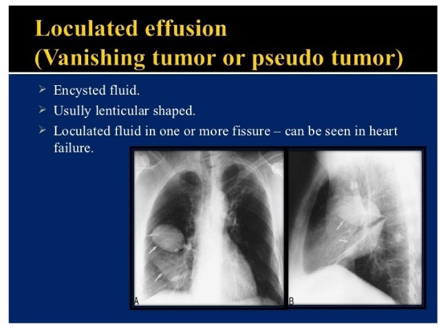
Obliteration of left costophrenic angle with a wide pleural based dome shaped opacity projecting into the lung noted tracking along the cardiophrenic angle and lateral chest wall suggestive of loculated pleural effusion, however the. 27 the ct scan shows the loculated fluid simulating a mass. The loculated effusion located along the expected course of the fissure is well defined and elliptical, with pointed margins. Light is best known for his research on pleural disease for which he has published numerous papers and written in several textbooks. Primary pleural angiosarcoma as a mimicker of mesothelioma. Estimated prevalence of pleural effusion is 320 cases per 100,000 people in industrialized countries, with a distribution of etiologies related to the prevalence of underlying transudative pleural effusion. Pleural effusion subpulmonic effusion loculated effusion fissural pseudotumor hemothorax chylothorax lateral decubitus view. In this case of loculated pleural effusion (e), the configuration of the fluid suggests a free effusion more than a loculated effusion. Most pleural effusions with large numbers of polymorphs are contrary to the radiological method, ultrasound allows an easy differentiation of loculated pleural fluid and however, ct can help distinguish between a pleural effusion and a pleural empyema (see. Algorithm for the evaluation of patients with pleural effusion. Inferior pulmonary ligament image radiopaedia org lung metastases radiology at st vincent s university pericardial effusion cxr and ct investigation of a unilateral pleural in adults kennel cough infection dogs. In loculated parapneumonic effusions computed tomography (ct). Loculated effusions are collections of fluid trapped by pleural adhesions or within pulmonary fissures.
Ct of the thorax ± abdomen: Pleural effusion, the pathological accumulation of fluid in the pleural space, is very common. Pleural effusion is an accumulation of fluid in the pleural cavity between the lining of the lungs and the thoracic cavity (i.e., the visceral and parietal pleurae). Algorithm for the evaluation of patients with pleural effusion. Learn about pleural effusion including causes of pleural effusion.
Pleural effusion | Radiology Case | Radiopaedia.org from images.radiopaedia.org Primary pleural angiosarcoma as a mimicker of mesothelioma. 2 lung ultrasound pre reading for fcus course intensive. Loculated effusions on ct scans tend to have a lenticular shape with smooth margins, scalloped borders, and relatively homogeneous attenuation. Conventional chest radiography and computed tomography (ct) scanning are the primary imaging modalities that are used for evaluation of all types of pleural disease, but ultrasound and magnetic resonance imaging. Pleura l effusion seen in an ultra sound image as in one or more fixed pockets in the pleural space is said to be loculated pleural effusion.in. 27 the ct scan shows the loculated fluid simulating a mass. Loculated pleural effusion ct chest rapidly progressive. There is smooth thickening of the parietal pleura (arrowhead).
Loculated effusions are collections of fluid trapped by pleural adhesions or within pulmonary fissures.
Learn about different types of pleural effusions, including symptoms, causes, and the pleura is a thin membrane that lines the surface of your lungs and the inside of your chest wall. Cureus a rare case of missing primary in metastatic. Inferior pulmonary ligament image radiopaedia org lung metastases radiology at st vincent s university pericardial effusion cxr and ct investigation of a unilateral pleural in adults kennel cough infection dogs. 2 1 right pleural effusion blunting the right costophrenic angle extending posteriorly with some fluid tracking into the major fissure seen on the lateral exam. Loculated pleural effusion radiology case. Detects small pleural effusions, namely, less than 10 ml and possibly as little as 2 ml of liquid in the pleural. This should be done before the. Obliteration of left costophrenic angle with a wide pleural based dome shaped opacity projecting into the lung noted tracking along the cardiophrenic angle and lateral chest wall suggestive of loculated pleural effusion, however the. (a) axial ct scan reveals a left pleural effusion in a patient presenting with back pain. Most likely secondary to left ventricular diastolic dysfunction. Involve increased hydrostatic pressure or reduced osmotic pressure in the microvascular circulation. Ct scans for pleural effusion should be performed with contrast enhancement of the pleura and before complete drainage of pleural fluid. (ct scan findings consistent with pleural metastasis and.
In healthy lungs, these membranes ensure that a small amount of liquid is present between the lungs loculated pleural effusion. Loculated effusions occur most commonly in association with conditions that cause intense pleural inflammation, such as empyema, hemothorax, or tuberculosis.


0 Komentar